




3D Mammography
UHS can provide you with the latest 3D technology for breast cancer screening
As part of our ongoing commitment to you, we are proud to offer 3D mammography the latest in breast cancer screening, available right here at the UHS Breast Center in Vestal.
A 3D mammogram consists of multiple breast images taken in just seconds to produce a 3D image. The doctor looks through the tissue one millimeter at a time seeing detail in a way never before possible.
For women who undergo routine mammograms, 3D is another option and the latest diagnostic technology available.
To schedule your 3D mammogram in Vestal at the UHS Breast Center, call (607) 240-2847.
Images from a breast exam: 2D vs 3D Slices
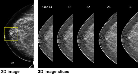
In a "conventional" 2D mammogram there appears to be an area of concern that the doctor may want to further investigate with another mammogram or biopsy. Looking at the same breast tissue in 3D mammography image slices, the doctor can now see that the tissue is in fact normal breast tissue that was overlapping in the traditional mammogram creating the illusion of an abnormal area. In this scenario this patient likely avoided a callback for an additional mammogram thanks to the 3D mammography exam technology.
Frequently Asked Questions about 3D Mammography
What is a 3D mammography breast exam?
3D mammography is a revolutionary new screening and diagnostic tool designed for early breast cancer detection that can be done in conjunction with a traditional 2D digital mammogram.
During the 3D part of the breast exam done in our Vestal office, the X-ray arm sweeps in a slight arc over your breast, taking multiple breast images. Then, a computer produces a 3D image of your breast tissue in one millimeter slices, providing greater visibility for the radiologist to see breast detail in a way never before possible. They can scroll through images of your entire breast like pages of a book.
The additional 3D images make it possible for a radiologist to gain a better understanding of your breast tissue during screening, significantly improving early breast cancer detection and providing the confidence to reduce the need for follow-up imaging by up to 40%.
Why is there a need for tomosynthesis breast exams? What are the benefits?
With conventional digital mammography, the radiologist is viewing all the complexities of your breast tissue in a one flat image. Sometimes breast tissue can overlap, giving the illusion of normal breast tissue looking like an abnormal area.
By also looking at the breast tissue in one millimeter slices, the radiologist can provide a more accurate exam. In this way, 3D mammography finds 40% more invasive cancer missed with conventional 2D mammography. It also means there is less chance your doctor will call you back later for a “second look,” because now they can see breast tissue more clearly.
What is the difference between a screening and diagnostic mammogram?
A screening mammogram is your annual mammogram that is done every year. Sometimes the radiologist may ask you to come back for follow-up images which is called a diagnostic mammogram to rule out an unclear area in the breast or if there is a breast complaint that needs to be evaluated.
What should I expect during the 3D mammography exam?
3D mammography complements standard 2D mammography and is performed at the same time with the same system. There is no additional compression required, and it only takes a few more seconds longer for each view.
Is there more radiation dose?
Very low X-ray energy is used during the exam, just about the same amount as a traditional mammogram done on film.
Who can have a 3D mammography exam?
It is approved for all women who would be undergoing a standard mammogram, in both the screening and diagnostic settings.
-
 Know where to go for your medical concernOctober 21, 2026
It can be tough to distinguish where to go for medical care when your symptoms feel unbearable, and your primary care provider is unavailable. Here are some key differences to help you decide.
-
 UHS Spine Surgery Program earns re-certification from Joint CommissionOctober 20, 2026
UHS has earned re-certification for the Joint Commission’s Gold Seal of Approval® for Core Certification in Spine Surgery by demonstrating continuous compliance with Joint Commission performance standards.
-
 UHS School-Based Nutrition Services is helping students build healthy habitsOctober 15, 2026
UHS Nutrition Services attended an after-school event for Supporting Our Minds and Bodies Through Wellness initiative at Benjamin Franklin Elementary School in Binghamton on October 8.
-
 Rain or Shine, Team UHS Makes StridesOctober 12, 2026
Rainy weather couldn’t dampen Team UHS’s spirit as staff members came together for this year’s Making Strides Against Breast Cancer walk at Mirabito Stadium in Binghamton on Sunday, October 11.